The Next Generation of Dental X-Rays
This technology allows the dentist to capture and reproduce the detailed 3D anatomy of the skull in a reliable, affordable way, while emitting low doses of radiation. Dr. Bedi is committed to providing his patients with the most advanced and safest technology available.
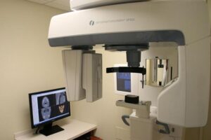 Cone beam imaging is one of the most important breakthroughs in dental radiology, and has proven effective for a wide range of care and treatment applications. Cone beam technology has several features that make high-resolution, 3D imaging a reality. A digital x-ray scanner is mounted on a rotating arm that circles the patient’s head. As it rotates, the x-ray is projected in a carefully controlled, cone-shaped beam through the patient and onto an amorphous silicon flat panel or image intensifier sensor. The beam encompasses the patient’s entire head, so it only takes one pass to capture the complete skull anatomy. The resulting images are displayed on a computer screen.
The Benefits:
Dramatically lower radiation emission: An average CT scan is about 600-700 microsieverts, while cone beam imaging is usually less than 70 microsieverts.
Short scan and reconstruction times: Scans take an average of about 20 seconds, and less than a minute later, images are reconstructed on the computer screen allowing to see—in 360°—undistorted, virtual, rotating models of the patient’s anatomy.
Better diagnosis: Bone thickness can be accurately measured to better determine implant candidates and better diagnosis of diseases or conditions.
The Cone Beam images have more intense details: Soft tissue, missing teeth, location of the nerve canals and the relationship between proposed implants and the opposite jaw are fully visible.
Call our Spring Hill, Fl dentist office today to schedule your consultation with Dr. Bedi. It is our goal to make you feel at home as part of the Anchor Dental Care family and answer any questions you may have about its practice and/or services.